Head & neck · Interactive 3D
Radial forearm flap
The radial forearm fasciocutaneous flap in three stages: marking and exposing the radial artery at the wrist, raising the paddle with its fascia and dissecting the pedicle proximally, and the harvested free flap with the radial artery, venae comitantes, cephalic vein and palmaris longus.
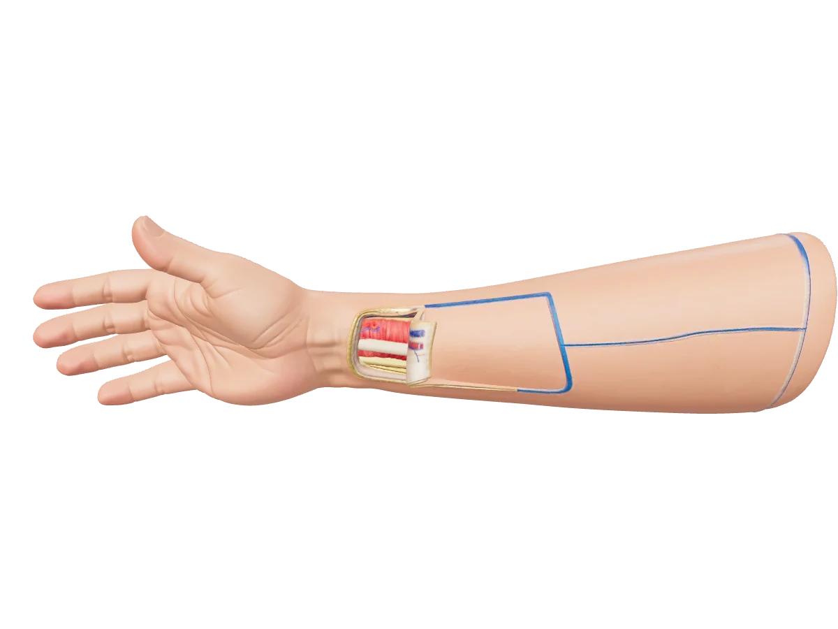
Step 1 · Mark
Mark the flap and expose the artery
Allen’s test, then mark the radial artery by its pulse, design the paddle with its radial edge over the artery, and open the distal end to find the vessels.
- Allen’s test first: the hand must perfuse through the ulnar artery and palmar arch once the radial artery is taken.
- The radial artery is palpable in the distal forearm; follow the pulse proximally and mark its course to the antecubital fossa.
- Design the paddle with its radial border over the artery, extending ulnarward onto the less hair-bearing skin; size it from a template of the defect.
- Shoulder abducted to 90° on an arm board, padded tourniquet proximally, and the distal end of the flap is opened first.
Hotspots in this step
- 1Radial artery at the wrist — Palpable between flexor carpi radialis and brachioradialis — the first structure found when the distal end of the flap is opened. Allen’s test before anything else.
- 2Venae comitantes — Paired veins with the artery; they can be small, which is why the cephalic vein is often taken as well.
- 3Skin paddle — Radial edge over the artery, extended ulnarward as needed; the paddle can be folded or split for three-dimensional defects.
- 4Pedicle marking — The course of the radial artery marked proximally — the pedicle can be dissected to its origin just distal to the antecubital fossa.
- 5Tourniquet — A padded tourniquet proximally; previous arterial or intravenous lines in this forearm compromise the flap and should be asked about.
Technique summary after microsurgeon.org (radial forearm flap).
More models
Anterolateral thigh (ALT) flap
The anterolateral thigh perforator flap raised on the descending branch of the lateral circumflex femoral artery, shown with the skin island lifted away from the thigh and the perforators entering it between rectus femoris and vastus lateralis.
Open modelBilobed flap (Zitelli)
An interactive 3D model of the Zitelli bilobed flap: one flap with two lobes at 45° and 90° pivoting on a single point, the primary lobe filling the defect, the secondary lobe filling the primary donor site, Burow's triangle at the pivot and direct closure of the secondary donor.
Open modelTemporal bone & middle ear
A right temporal bone opened to show the external canal, the tympanic membrane, the middle ear cleft with its medial wall — promontory, oval and round windows, the horizontal facial canal and the lateral semicircular canal — and the mastoid antrum and air cells behind.
PreviewStart with 20 free questions
Create a free account with Google and go straight into the sample bank, today's Daily Five and your readiness dashboard. No card required.