Basic science · Free article
Reading an audiogram: thresholds, masking and the patterns that matter
How to read a pure-tone audiogram for FRCS (ORL-HNS): BSA symbols and axes, degrees of hearing loss, the air–bone gap, the three masking rules and the classic patterns.
What the chart shows
Pure-tone audiometry measures the quietest tone a person can hear at each test frequency, in each ear, by air conduction through earphones and by bone conduction through a vibrator on the mastoid. The threshold is the level at which the tone is heard on at least half of its presentations. Frequency runs along the horizontal axis on a logarithmic scale, usually from 125 or 250 Hz to 8000 Hz, so each octave takes the same width. Hearing level runs down the vertical axis in decibels hearing level (dB HL), from −10 at the top to 120 at the bottom. 0 dB HL is the average threshold of young adults with normal hearing at that frequency, so a normal audiogram is a line near the top of the chart, and worse hearing plots lower.
The British Society of Audiology (BSA) recommends separate charts for the right and left ears, with the aspect ratio fixed at 20 dB to one octave so that the slope of a loss looks the same on every form. Octave frequencies are solid gridlines; the half-octaves (750, 1500, 3000 and 6000 Hz) are dashed and are tested when needed — 3000 and 6000 Hz in particular when there is a high-frequency loss. The usual test order is the better ear first, starting at 1000 Hz, then 2000, 4000, 8000, 500 and 250 Hz, with a retest at 1000 Hz in the first ear as a check of reliability.
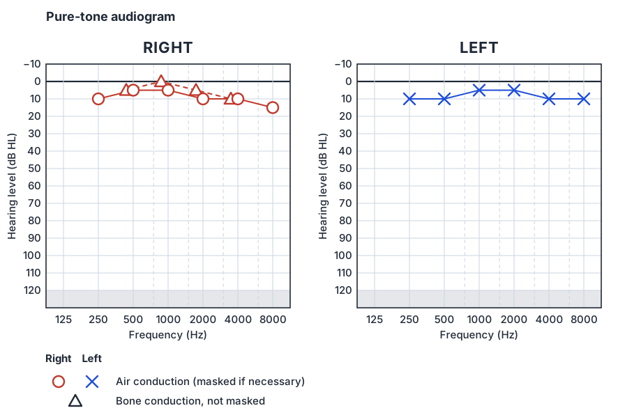
The symbols
| Measurement | Right | Left | Joined by |
|---|---|---|---|
| Air conduction (masked if necessary) | O | X | Continuous lines |
| Bone conduction, not masked | Δ (mastoid recorded) | Δ (mastoid recorded) | Broken lines |
| Bone conduction, masked | [ | ] | Broken lines |
| Uncomfortable loudness level | └ | ┘ | — |
| No response at the maximum output | Arrow pointing down from the corner of the symbol | Arrow pointing down from the corner of the symbol | Not joined |
Two details catch candidates out. First, the BSA air-conduction symbol means “masked if necessary”: a correctly completed audiogram shows true thresholds, and there is no separate masked air-conduction symbol. Other systems differ — the American (ASHA) symbols use a triangle and a square for masked air conduction — and audiology software varies, so always read the key. Second, the unmasked bone-conduction symbol belongs to neither ear. Because the skull carries bone-conducted sound to both cochleae with little or no loss, the Δ tells you only that the better cochlea heard the tone at that level.
Degree, type and configuration
Describe an audiogram in three steps: how much hearing is lost, what kind of loss it is, and what shape it has. The BSA descriptors are based on the average of the air-conduction thresholds at 250, 500, 1000, 2000 and 4000 Hz, with a no-response counted as 130 dB HL:
| Descriptor | Average hearing threshold (dB HL) |
|---|---|
| Mild | 21–40 |
| Moderate | 41–70 |
| Severe | 71–95 |
| Profound | Above 95 |
The type comes from comparing air with bone conduction. Air conduction tests the whole pathway — canal, drum, ossicles, cochlea and nerve. Bone conduction largely bypasses the outer and middle ear and tests the cochlea and beyond. So:
- Conductive: bone conduction normal, air conduction raised — an air–bone gap. Wax, effusion, perforation, ossicular fixation or discontinuity.
- Sensorineural: air and bone conduction raised together, with no gap. Cochlear or retrocochlear disease.
- Mixed: bone conduction raised, and air conduction raised further still — both elements at once.
Small differences of 5–10 dB between air and bone conduction are within the variability of the test and should not be over-read. Configuration completes the description: flat, sloping (worse in the high frequencies), rising (worse in the low frequencies), notched, or mid-frequency (“cookie-bite”).
Masking: who heard the tone?
When one ear hears much better than the other, a loud tone presented to the poorer ear can cross the skull and be heard by the better cochlea. The poorer ear then appears to have a threshold — a “shadow” of the better ear — that is not its own. The loss in crossing the head is the interaural attenuation. The BSA gives it as 40–80 dB with supra-aural or circum-aural earphones, at least 55 dB with correctly inserted insert earphones, and little or nothing for bone conduction. Masking puts a narrow-band noise into the non-test ear to occupy it while the test ear is measured.
The BSA sets out three indicators of cross-hearing, applied frequency by frequency:
- Rule 1: the unmasked air-conduction thresholds of the two ears differ by 40 dB or more (55 dB or more with insert earphones). Mask the better ear while retesting the worse ear by air conduction.
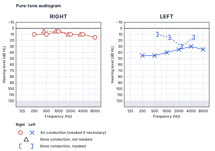
- Rule 2: the unmasked bone-conduction threshold is 10 dB or more better than the air-conduction threshold of either ear. The worse ear by air conduction is the test ear, and the better ear is masked to find its true bone-conduction threshold.
- Rule 3: where Rule 1 did not apply, the bone-conduction threshold of one ear is 40 dB or more (55 dB with inserts) better than the unmasked air-conduction threshold of the other. This catches cross-hearing when the non-test ear has a conductive loss of its own.
The figure shows why it matters. The left ear has an air–bone gap, so Rule 2 applies, and bone conduction has been masked: the brackets show the left cochlea is near normal, so the loss is conductive. Without masking, the Δ alone could not have told you which cochlea was responding.
Patterns worth recognising
- Presbyacusis
- Bilateral, roughly symmetrical sensorineural loss, sloping down towards the high frequencies, with no air–bone gap.
- Noise exposure
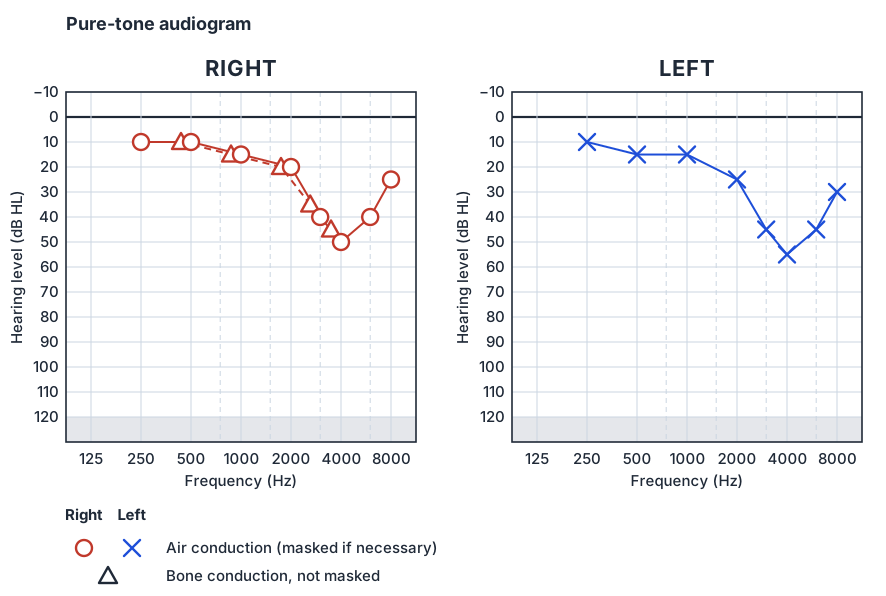
- A sensorineural notch at 3–6 kHz, often deepest at 4 kHz, with some recovery at 8 kHz — usually bilateral, though a shooter’s ear may be worse on one side.
- Otosclerosis
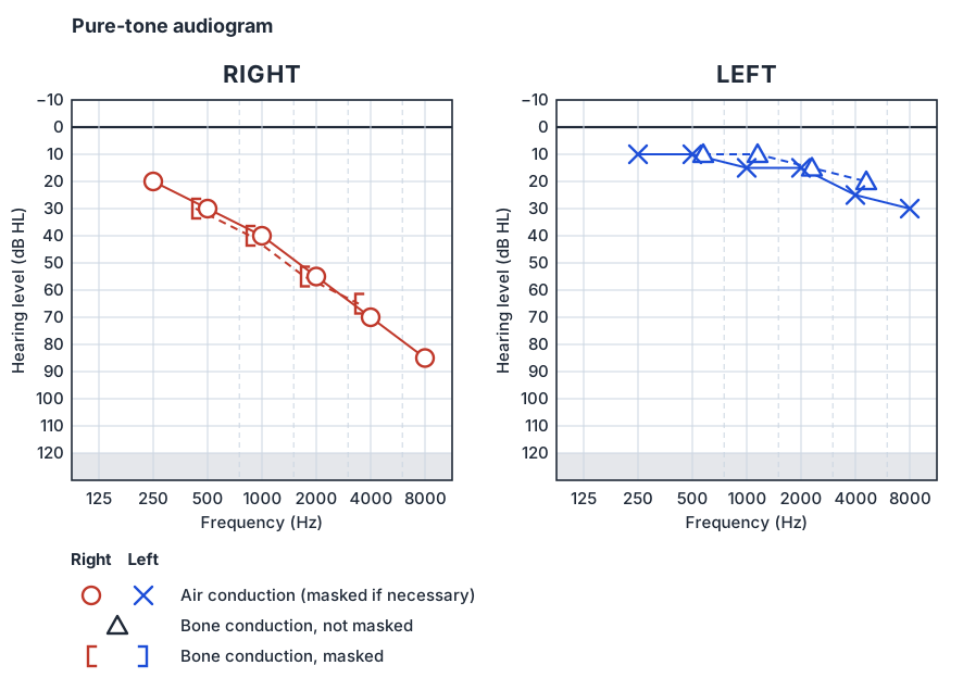
- A conductive loss, often greater in the low frequencies, with a dip in bone conduction around 2 kHz (Carhart’s notch). The notch is a mechanical effect of stapes fixation and improves after successful stapes surgery.
- Otitis media with effusion
- A mild, fairly flat conductive loss with a flat tympanogram and a normal ear-canal volume.
- Ménière’s disease
- A low-frequency sensorineural loss that fluctuates in the early stages, with episodic vertigo, tinnitus and aural fullness.
- Ototoxicity
- Bilateral, high-frequency sensorineural loss that progresses downwards with the cumulative dose, typically after cisplatin.
- Asymmetric sensorineural loss
- One ear worse than the other with no conductive cause. NICE suggests considering MRI of the internal auditory meati for an asymmetry of 15 dB or more at two adjacent test frequencies (0.5, 1, 2, 4 and 8 kHz), and offering it whenever there are localising symptoms or signs.
Traps and artefacts
- The unmasked bone-conduction symbol. It cannot be attributed to an ear. A conductive loss in the worse ear is only proved by masked bone conduction.
- Vibrotactile responses. At high levels and low frequencies a bone vibrator can be felt rather than heard, producing an apparent air–bone gap in a profoundly deaf ear.
- Collapsing ear canals. Supra-aural earphones can close a soft canal and create a spurious high-frequency conductive loss; insert earphones avoid it.
- Non-organic hearing loss. Thresholds that vary between tests, or a poor ear with no shadow curve where cross-hearing should have occurred, should prompt objective testing.
- A false-negative Rinne. A dead ear can give a negative Rinne because the opposite cochlea hears the bone-conducted tone. The audiogram, with masking, resolves it.
Frequently asked questions
What do O and X mean on an audiogram?
O is the right ear and X the left ear by air conduction, masked where necessary. In British Society of Audiology format, Δ is bone conduction not masked, and [ and ] are masked bone conduction for the right and left ears.
When is masking needed in pure-tone audiometry?
When cross-hearing is possible. The BSA rules are: an air-conduction difference between the ears of 40 dB or more (55 dB with insert earphones); an unmasked bone-conduction threshold 10 dB or more better than the air-conduction threshold of either ear; and a bone-conduction threshold in one ear 40 dB (55 dB with inserts) better than the unmasked air-conduction threshold of the other.
How is the degree of hearing loss described?
The BSA uses the average air-conduction threshold at 250, 500, 1000, 2000 and 4000 Hz: 21–40 dB HL is mild, 41–70 moderate, 71–95 severe and above 95 profound.
What is an air–bone gap?
The difference between the air-conduction and bone-conduction thresholds at a frequency. A gap with normal bone conduction means a conductive loss; small gaps of 5–10 dB are within the variability of the test.
What does a 4 kHz notch mean?
A sensorineural dip at 3–6 kHz, deepest at 4 kHz and recovering at 8 kHz, is the typical audiometric pattern of noise damage.
References
- British Society of Audiology. Recommended procedure: pure-tone air-conduction and bone-conduction threshold audiometry with and without masking. 2018.
- NICE. Hearing loss in adults: assessment and management (NG98). 2018.
- Carhart R. Clinical application of bone conduction audiometry. Arch Otolaryngol 1950;51(6):798–808.
- Watkinson JC, Clarke RW (eds). Scott-Brown’s Otorhinolaryngology and Head and Neck Surgery. 8th ed. CRC Press; 2018.
Further reading
- British Society of Audiology recommended procedures — The current procedures for pure-tone audiometry, masking and tympanometry
- NICE NG98: hearing loss in adults — Referral and imaging criteria, including the asymmetry threshold for MRI
Written for revision in the FRCS (ORL-HNS) style under our editorial standards. It is not clinical advice: always confirm against current guidance. Spotted an error? Tell us.
Related articles
Basic science
Tympanometry: reading a tympanogram
How to read a tympanogram for FRCS (ORL-HNS): peak pressure, peak admittance and ear-canal volume, BSA normal ranges, Jerger types, flat traces and the 1000 Hz infant test.
ReadOtology
Sudden sensorineural hearing loss: recognition, referral and treatment
Sudden sensorineural hearing loss for FRCS (ORL-HNS): the definition, NICE referral timings, ruling out a conductive cause, MRI, steroids, salvage treatment and follow-up.
ReadOtology
The facial nerve: anatomy, localisation and House–Brackmann grading
The facial nerve for FRCS (ORL-HNS): its course from brainstem to face, the branches that localise a lesion, House–Brackmann grades I–VI, Bell’s palsy and the eye.
ReadTurn reading into marks
Drill basic science with curriculum-mapped questions, rate your confidence, and watch the gap close on your dashboard.