Basic science · Free article
Tympanometry: reading a tympanogram
How to read a tympanogram for FRCS (ORL-HNS): peak pressure, peak admittance and ear-canal volume, BSA normal ranges, Jerger types, flat traces and the 1000 Hz infant test.
What tympanometry measures
Tympanometry is an objective test of the middle ear, not of hearing. A probe sealed in the ear canal plays a low-frequency tone — 226 Hz in children over about six months and adults — and measures how much of it the system accepts, its admittance, while a pump sweeps the air pressure in the canal from positive to negative. The eardrum moves most freely when the pressure on its two sides is equal, so admittance peaks when canal pressure matches middle-ear pressure. Away from that point the drum is pushed or pulled taut and admittance falls. At 226 Hz the units ml, cm³ and mmho are interchangeable, which is why the vertical axis is often labelled in ml.
The BSA recommends a slow sweep, starting at +200 daPa and continuing to about −300 daPa, going further (to −600 daPa) when searching for a peak in a very negative ear. Descending, slower sweeps also reduce the tendency of tympanometry to overestimate the ear-canal volume.
The three numbers
| Measurement | What it is | BSA reference values |
|---|---|---|
| Tympanic peak pressure (TPP) | The canal pressure at which the peak occurs — an estimate of middle-ear pressure | −50 to +50 daPa can be considered normal in adults; down to −100 daPa may be of little significance on its own; in children a TPP as low as −150 daPa may be of little clinical significance |
| Peak admittance (Ytm, often called compliance) | The height of the peak with the ear canal’s contribution removed | 0.3–1.6 mmho in 90% of adults; 0.2 mmho is an acceptable lower limit from 6 months to 6 years; 0.2–1.0 mmho in 90% of children aged 5–7 |
| Ear-canal volume (ECV) | The admittance of the air in front of the probe, read where the drum is stiff at the pressure extremes | 0.6–2.2 cm³ in 90% of adults (larger in men); about 0.6 cm³ in infants; 0.7–1.3 cm³ in 90% of children aged 5–7 |
The BSA makes two further points worth knowing for the exam. Tympanic peak pressure overestimates true middle-ear pressure, by 30–70 daPa in some circumstances, and is a snapshot: a single negative reading suggests Eustachian tube dysfunction but does not prove it. And the effect of negative pressure on hearing is better judged from the low-frequency air–bone gap on the audiogram than from the pressure itself.
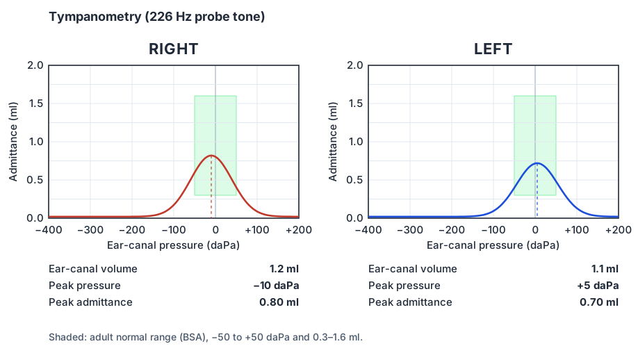
Jerger types — and why the numbers matter more
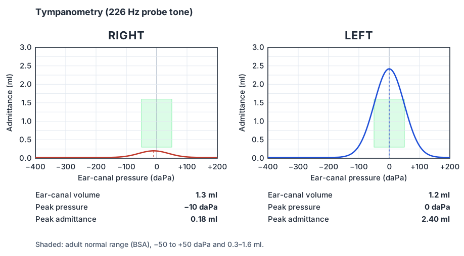
Jerger’s 1970 classification describes the shape of the trace, and it is still how many clinicians and many exam questions talk about tympanograms:
- Type A
- A normal peak at normal pressure.
- Type As
- A shallow peak at normal pressure: a stiff system, as in otosclerosis or a thickened, scarred drum.
- Type Ad
- A deep, tall peak at normal pressure: a hypermobile system, as in ossicular discontinuity or a thin, flaccid segment of the drum.
- Type B
- No peak — a flat trace. Effusion, perforation, a patent grommet, wax or a probe against the canal wall; the ear-canal volume tells them apart.
- Type C
- A peak at negative pressure: Eustachian tube dysfunction or a resolving effusion.
The flat trace
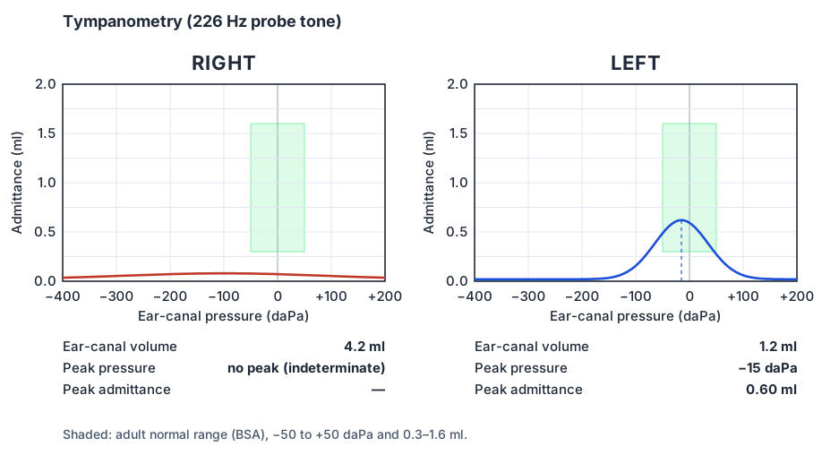
A flat tympanogram has no peak, so the middle-ear pressure cannot be measured. Its meaning depends on the ear-canal volume, which is why the ECV must always be read with a flat trace — and compared between the two ears:
- Normal ECV: the probe sees a normal canal ending at a drum that will not move — middle-ear effusion is the usual cause.
- Large ECV: the probe sees the canal plus the middle ear and mastoid through an open hole — a perforation or a patent grommet. The BSA notes that a volume of 3.6 ml or more that differs from the other ear may be due to a perforation.
- Small ECV: the probe is against the canal wall or blocked by wax or debris. Look in the ear and repeat the test.
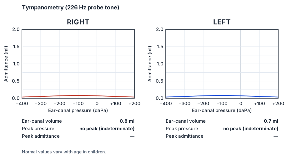
In children with suspected otitis media with effusion, NICE NG233 asks for formal assessment including tympanometry and age-appropriate audiometry, and for hearing to be reassessed after three months in bilateral OME with hearing loss. Tympanometry shows the effusion; the audiogram shows what it is doing to hearing, and that is what decides management.
Infants and the 1000 Hz probe
In babies the ear canal walls are soft and the middle ear behaves differently, so a 226 Hz tympanogram can look normal even when there is fluid. A 1000 Hz probe tone is used instead below 6 months of corrected age. The BSA recommends classifying these traces as normal or abnormal with Baldwin’s method: draw a baseline between the pressure extremes; a positive peak above the baseline is normal and a negative peak (a trough) is abnormal. The ear-canal volume is disregarded at 1000 Hz because it is not precise, except as a clue that the probe is blocked.
Acoustic reflexes
The same probe can measure the stapedial reflex — a small drop in admittance when a loud sound makes stapedius contract. The reflex arc runs from the cochlea through the eighth nerve to the brainstem and back through the facial nerve on both sides, so the pattern of present and absent ipsilateral and contralateral reflexes can localise a lesion. Reflexes are absent with a conductive loss in the probe ear, may be present at reduced sensation levels in a cochlear loss with recruitment, and may be absent or decay in retrocochlear disease. In practice MRI has replaced reflex testing for detecting vestibular schwannoma, but the principles still appear in questions.
Frequently asked questions
What is a normal tympanogram?
A single sharp peak near 0 daPa. The BSA gives −50 to +50 daPa as normal middle-ear pressure in adults, 0.3–1.6 ml as the 90% range for adult peak admittance and 0.6–2.2 cm³ for adult ear-canal volume.
What does a type B tympanogram with a large ear-canal volume mean?
A flat trace with a large canal volume suggests the probe is measuring the middle ear as well as the canal through an opening: a perforation or a patent grommet. With a normal canal volume, a flat trace suggests effusion; with a small one, a blocked probe.
What does a type C tympanogram mean?
A peak at negative pressure, suggesting Eustachian tube dysfunction or a resolving effusion. The BSA notes that pressures down to −100 daPa may be of little significance in adults on their own, and down to −150 daPa in children.
Why is a 1000 Hz probe used in young babies?
Because the ear canal and middle ear of a young baby behave differently, and a 226 Hz trace can look normal even when there is fluid. Below 6 months of corrected age the BSA recommends a 1000 Hz probe, with the trace read as a positive or negative peak against a baseline.
What does tympanometry show in otosclerosis?
Usually a peak at normal pressure, sometimes with low peak admittance (Jerger type As), and absent acoustic reflexes in the affected ear. A normal tympanogram with a conductive loss on the audiogram is the classic pairing. Ossicular discontinuity, by contrast, can give an unusually high peak (type Ad).
References
- British Society of Audiology. Recommended procedure: tympanometry and acoustic reflex thresholds. February 2024, minor amendments February 2025, March 2026 and November 2026.
- Jerger J. Clinical experience with impedance audiometry. Arch Otolaryngol 1970;92(4):311–324.
- NICE. Otitis media with effusion in under 12s (NG233). 2023.
- Watkinson JC, Clarke RW (eds). Scott-Brown’s Otorhinolaryngology and Head and Neck Surgery. 8th ed. CRC Press; 2018.
Further reading
- BSA recommended procedure: tympanometry and acoustic reflex thresholds — Normal values, reporting and the 1000 Hz method in full
Written for revision in the FRCS (ORL-HNS) style under our editorial standards. It is not clinical advice: always confirm against current guidance. Spotted an error? Tell us.
Related articles
Basic science
Reading an audiogram: thresholds, masking and the patterns that matter
How to read a pure-tone audiogram for FRCS (ORL-HNS): BSA symbols and axes, degrees of hearing loss, the air–bone gap, the three masking rules and the classic patterns.
ReadOtology
Sudden sensorineural hearing loss: recognition, referral and treatment
Sudden sensorineural hearing loss for FRCS (ORL-HNS): the definition, NICE referral timings, ruling out a conductive cause, MRI, steroids, salvage treatment and follow-up.
ReadTurn reading into marks
Drill basic science with curriculum-mapped questions, rate your confidence, and watch the gap close on your dashboard.